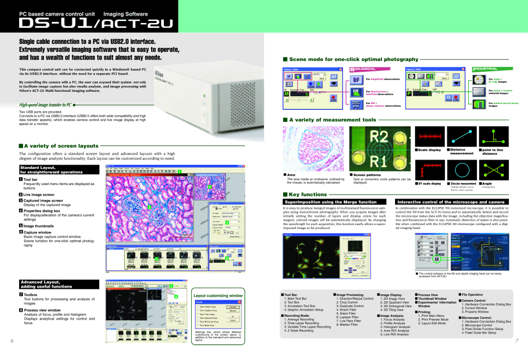
PC based camera control unit Imaging Software
Single cable connection to a PC via USB2.0 interface. Extremely versatile imaging software that is easy to operate, and has a wealth of functions to suit almost any needs.
This compact control unit can be connected quickly to a Windows® based PC via its USB2.0 interface, without the need for a separate PCI board.
By controlling the camera with a PC, the user can expand their system, not only to facilitate image capture but also results analysis, and image processing with Nikon's
Two USB ports are provided.
Connects to a PC via USB2.0 interface (USB2.0 offers both wide compatibility and high data transfer speeds), which enables camera control and live image display at high speed on a monitor.
 A variety of screen layouts
The configuration offers a standard screen layout and advanced layouts with a high degree of image analysis functionality. Each layout can be customized according to need.
 Scene mode for
 one-click optimal photography
For brightfield observations |
|
|
|
| For wafer / |
|
|
|
|
| IC chip images |
For fluorescence / |
|
|
| For metal / ceramic | |
darkfield observations |
| material images | |||
|
| ||||
For DIC / |
| For printed circuit board | |||
phase contrast observations |
| images | |||
 A variety of measurement tools
| Scale display |
| Distance |
| point to line |
|
|
| measurement |
| distance |
Standard Layout,
for straightforward operations
1Tool bar
Frequently used menu items are displayed as buttons
2Live image screen
3Captured image screen Display of the captured image
4Properties dialog box
For display/alteration of the camera’s current settings
5Image thumbnails
6Capture window
Basic image capture control window
Scene function for
![]() 1
1
2
3
4
Area |
| Screen patterns |
|
|
|
|
|
|
|
|
|
|
|
|
|
|
|
|
|
|
|
|
|
|
|
|
|
|
|
|
|
|
| ||
|
|
|
|
|
|
|
|
|
|
|
|
|
|
|
| ||
|
|
|
|
|
|
|
|
|
|
|
|
|
|
|
| ||
|
|
|
|
|
|
|
|
|
|
|
|
|
|
|
| ||
|
|
|
|
|
|
|
|
|
|
|
|
|
|
|
| ||
|
|
|
|
|
|
|
|
|
|
|
|
|
|
|
| ||
The area inside an enclosure, outlined by |
| Grid or concentric circle patterns can be |
|
|
|
|
|
|
|
|
|
|
|
|
|
|
|
the mouse, is automatically calculated |
| displayed. |
| XY scale display |
| Circular measurement |
| Angle | |||||||||
|
|
|
| ||||||||||||||
|
|
|
|
|
|
|
|
|
|
|
|
|
| (displays diameter, circum- |
| (intersection) | |
|
|
|
|
|
|
|
|
|
|
|
|
|
| ference, center of gravity) |
|
| |
 Key functions
Superimposition using the Merge function |
| Interactive control of the microscope and camera |
It is easy to produce merged images of multistained fluorescence sam- |
| In combination with the ECLIPSE 90i motorized microscope, it is possible to |
ples using monochrome photography. When you acquire images after |
| control the 90i from the |
initially setting the number of layers and display colors for each |
| the microscope status data with the image, including the objective magnifica- |
reagent, colored images will be automatically displayed. By changing |
| tion and fluorescence filter in use. Automatic detection of status is also possi- |
the wavelength for each acquisition, this function easily allows a super- |
| ble when combined with the ECLIPSE 80i microscope configured with a digi- |
imposed image to be produced. |
| tal imaging head. |
Advanced Layout, adding useful functions
7Toolbox
Tool buttons for processing and analysis of images
8Process view window
Analysis of focus, profile and histogram Displays analytical settings for control and focus
6
5 |
| 6 |
|
![]() 7
7
8Layout customizing window
Settings box, which allows detailed customizing of the screen layout, in addition to the standard and advanced layout.
![]() The control software of the 90i and digital imaging head can be easily accessed from
The control software of the 90i and digital imaging head can be easily accessed from
Tool Bar: |
| Image Processing: |
|
| Image Display: |
| Process View |
| File Operation | |
|
|
|
|
| ||||||
1. Main Tool Bar |
| 1. Direction/Resize Control | 1. | 2D Image View |
| Thumbnail Window |
| Camera Control: | ||
2. Tool Box |
| 2. Crop Control | 2. | 2D Quadrant View |
| Experimental Information |
| 1. Hardware Connection Dialog Box | ||
3. Annotation Tool Box |
| 3. Duplicate Control | 3. | 3D Orthogonal View |
| Window |
| |||
|
|
| 2. Control Window | |||||||
4. Graphic Annotation Setup |
| 4. Kirsch Filter | 4. | 3D Tiling View |
|
|
| |||
|
| Printing: |
| 3. Property Window | ||||||
Recording Mode: |
| 5. Sobel Filter |
|
|
|
|
|
| ||
|
|
| Image Analysis: |
| 1. Print Main Menu |
|
| |||
1. Average Recording |
| 6. Laplace Filter |
|
| 1. Focus Analysis |
| 2. Print Preview Mode |
| Microscope Control: | |
| 7. Low Pass Filter |
|
|
|
| 1. Hardware Connection Dialog Box | ||||
2. Time Lapse Recording |
| 2. | Profile Analysis |
| 3. Layout Edit Mode |
| ||||
| 8. Median Filter |
|
| 2. Microscope Control | ||||||
3. Variable Time Lapse Recording |
| 3. | Histogram Analysis |
|
|
| ||||
|
|
|
|
| 3. Pixel Divide Function Setup | |||||
4. Z Stack Recording |
|
|
|
| 4. Area ROI Analysis |
|
|
| ||
|
|
|
|
|
|
| 4. Fixed Scale Bar Setup | |||
|
|
| 5. | Line ROI Analysis |
|
|
| |||
|
|
|
|
|
|
| ||||
7