9.Rotate the lens turret (item #10) to select the right lens for ideal viewing.

10.The greater the magnification the darker the room should be.

HORIZONTAL PROJECTION

1.Holding the neck of the microprojector with your left hand, place your right hand around the back of the dust proof revolving disk (item #14 avail- able on X-1000-1 only) and pull the projecting mechanism up into the hori- zontal position

2.When using liquids and projecting horizontally, the instrument head must be placed in a vertical position. Bring the specimen into focus while in the vertical position, then move the mirror (item# 15) into the light path and adjust so the image appears on the screen. Select the lens desired and refocus.

LIGHT POLARIZATION

1.Rotate the polarizing lens/analyzer (item #6 - white) until the lens is beneath the light beam.

2.Rotate the dust proof revolving disk (item #14) until the RED “P” on the edge of the disk is directly below the RED line.

3.Place a slide containing polarizing material on the stage.

4.Bring into focus and change colors by turning the polarizing lens/analyz- er. (item #5)

USE AS A MICROSCOPE

(X-1000-1 only)

1.Rotate the auxiliary lens disk (item #14) until the RED “E” on the edge of the disk is directly below the RED line.

2.Rotate the light diffusing lens (item #5-darker lens) until it is under the light beam.

3.Place the unit in the horizontal position.

4.View the specimen through the 10X eyepiece tube (item #16).

5.Bring into focus using the 16mm lens; then select the lens desired.

MODEL TRIG

OPERATING INSTRUCTIONS

Instructions covering the use of Ken-A-Vision Microprojector Model X1000-

1 apply except for the following:

1.Under “Vertical Projection” instructions disregard #4. Model Trig has only one objective lens.

2.Under “Vertical Projection” instructions disregard #7 entirely. Turning the knob between the two mounting rods adjusts the focus.

3.Under “Vertical Projection” disregard #9 in its entirety.

4.Instructions for the light polarization and use as a microscope are the same as for Model X1000-1

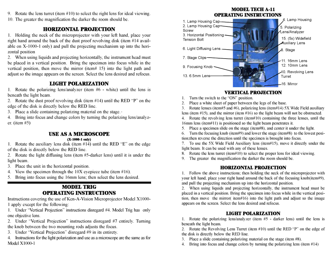
MODEL TECH A-11


OPERATING INSTRUCTIONS

1.

Lamp Housing Cap

4. Lamp Housing


2.

Lamp Housing Cap

5. Polarizing

Screw

Lens/Analyzer

3.

Horizontal Positioning

15. (5x) Widefield

Tension Bolt


Auxiliary Lens

6.

Light Diffusing Lens

8. Stage


7.

Stage Clips

11. 16mm Lens


9.

Focusing Knob

12. 10mm Lens


13. 6.5mm Lens

10. Revolving Lens

Turret

16. Mirror

VERTICAL PROJECTION

1.Turn the switch to the “ON” position.

2.Place a white sheet of paper between the legs of the base.

3.Rotate lenses (item#5 and #6), polarizing lens (item#14) 5X Wide Field auxiliary lens (item #15), and the mirror (item #16) so the light beam will not be obstructed.

4.Rotate the revolving lens turret (item#10) containing the three lenses, until the 16mm lens (item#11) is positioned so the light beam penetrates it.

5.Place a specimen slide on the stage (item#8), and center it under the light.

6.Turn the focusing knob (item#9) and lower the stage (item#8) to the lowest posi- tion;then reverse the direction until the specimen is brought into focus.

7.To use the 5X Wide Field Auxiliary lens (item#15), move it directly under the light beam. It can be used with any of these lenses.

8.Rotate the lens turret (item#10) to select the proper lens for ideal viewing.

9.The greater the magnification the darker the room should be.

HORIZONTAL PROJECTION

1.Follow the above instructions; then holding the neck of the microprojector with your left hand, place your right hand around the back of the focusing knob(item#9), and pull the projecting mechanism up into the horizontal position.

2.When using liquids and projecting horizontally, the instrument head must be placed in a vertical position. Bring the specimen into focus while in the vertical posi- tion, then move the mirror( item#16) into the light path and adjust so the image appears on the screen. Select the lens desired and refocus.

LIGHT POLARIZATION

1.Rotate the polarizing lens/analyzer (item #5 - darker lens) until the lens is beneath the light beam.

2.Rotate the Revolving Lens Turret (item #10) until the RED “P” on the edge of the disk is directly below the RED line.

3.Place a slide containing polarizing material on the stage (item #8).

4.Bring into focus and change colors by turning the polarizing lens (item #14)

Page 1
Image 1
Ken-A-Vision X1000 operating instructions Horizontal Projection, Light Polarization, USE AS a Microscope

X1000 specifications

Ken-A-Vision X1000 is an advanced educational microscope that integrates state-of-the-art technology, designed to enhance the learning experience in both classroom and laboratory settings. With its robust features and user-friendly interface, the X1000 is ideal for students, educators, and professionals alike.

One of the most notable features of the Ken-A-Vision X1000 is its high-resolution imaging capability. It utilizes a premium 5 megapixel camera, allowing users to capture crystal-clear images and videos of specimens in real time. The camera is paired with advanced optical components, providing excellent light transmission and clarity for detailed observations, making it perfect for both biological and materials science applications.

Equipped with LED illumination, the X1000 offers adjustable brightness levels, ensuring optimal viewing conditions for various types of samples. This feature not only prolongs the life of the light source but also reduces eye strain during extended use, enhancing the comfort of the user over lengthy viewing sessions.

The Ken-A-Vision X1000 is designed with versatility in mind. It includes multiple magnification options ranging from 40x to 1000x, which can be easily selected through a rotating turret. This flexibility allows users to switch between low and high magnification quickly, facilitating comprehensive examinations of specimens from different perspectives.

Another significant characteristic of the X1000 is its compatibility with various digital tools and applications. It supports plug-and-play functionality with simple USB connectivity to computers and laptops, enabling users to easily transfer images and videos for documentation or further analysis. This feature supports a wide range of educational software applications, making it an ideal choice for digital learning environments.

The microscope is also built with durability in mind. Its sturdy construction can withstand the rigors of frequent use in educational institutions while maintaining performance reliability. Moreover, the sleek and ergonomic design of the X1000 ensures ease of handling, allowing for comfortable operation without compromising functionality.

In summary, the Ken-A-Vision X1000 stands out as a premier educational microscope, combining high-resolution imaging, adjustable LED illumination, versatile magnification options, and digital compatibility into a user-friendly, durable design. Whether in a classroom or research facility, the X1000 is equipped to support and enhance the study of microscopic life and materials, making it an invaluable tool for exploration and discovery.