OPTICAL TERMINOLOGY
•Total magnification on the video monitor:
m(ob)=50⋅ , M(video camera adapter) is 0.5⋅ from Figure 1 and monitor magnification is 46.2⋅ from Figure 3.
M(monitor observation)=M(ob)⋅ M(video camera adapter)⋅ monitor magnification
=50⋅ 0.5⋅ 46.2=1155⋅
•Practical filed of view for video observation(horizontal side):
M(ob)=50⋅ , M(video camera adapter) is 0.5⋅ from Figure 1 and
horizontal side of 2/3" imaging device is 8.8mm from Figure 2
Practical field of view | = | Image device size |
for video observation |
| M(ob) ⋅ M(video camera adapter) |
8.8 (mm) |
|
|
= 50 ⋅ 0.5 =352µm
6. Numerical Aperture (N.A.)
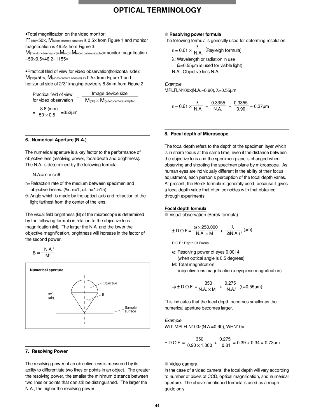
The numerical aperture is a key factor to the performance of objective lens (resolving power, focal depth and brightness). The N.A. is determined by the following formula:
N.A.= n ⋅ sinθ
n=Refraction rate of the medium between specimen and objective lenses. (Air: n=1, oil: n=1.515)
θ: Angle which is made by the optical axis and refraction of the light farthest from the center of the lens.
The visual field brightness (B) of the microscope is determined by the following formula in relation to the objective lens magnification (M). The larger the N.A. and the lower the objective magnification, brightness will increase in the factor of the second power.
B ∝ | N.A.2 | |
M2 | ||
|
Numerical aperture
Objective
n=1 | θ |
(air) |
|
Sample surface
7. Resolving Power
The resolving power of an objective lens is measured by its ability to differentiate two lines or points in an object. The greater the resolving power, the smaller the minimum distance between two lines or points that can still be distinguished. The larger the N.A., the higher the resolving power.
●Resolving power formula
The following formula is generally used for determing resolution.
ε = 0.61 ⋅ | λ | (Reyleigh formula) |
N.A. |
λ: Wavelength or radiation in use
(λ =0.55µm is used for visible light) N.A.: Objective lens N.A.
Example
MPLFLN100⋅ (N.A.=0.90), λ =0.55µm
ε = 0.61 ⋅ | λ | 0.3355 | 0.3355 |
| ||
| = |
| = |
| = 0.37µm | |
N.A. | N.A. | 0.90 | ||||
8. Focal depth of Microscope
The focal depth refers to the depth of the specimen layer which is in sharp focus at the same time, even if the distance between the objective lens and the specimen plane is changed when observing and shooting the specimen plane by microscope. As human eyes are individually different in the ability of their focus adjustment, each person's perception of the focal depth varies. At present, the Berek formula is generally used, because it gives a focal depth value that often coincides with that obtained through experiments.
Focal depth formula
●Visual observation (Berek formula)
| ω ⋅ 250,000 |
| λ | (µm) | |
|
|
|
| ||
± D.O.F.= N.A. ⋅ M | + | 2(N.A.) 2 | |||
| |||||
D.O.F.: Depth Of Focus
ω: Resolving power of eyes 0.0014 (when optical angle is 0.5 degrees)
M:Total magnification
(objective lens magnification x eyepiece magnification)
➔ ± D.O.F. = | 350 | + | 0.275 | (λ =0.55µm) |
N.A. ⋅ M | 2 | |||
|
| N.A. | ||
This indicates that the focal depth becomes smaller as the numerical aperture becomes larger.
Example
With MPLFLN100⋅ (N.A.=0.90), WHN10⋅ :
3500.275
± D.O.F. = 0.90 ⋅ 1,000 + 0.81 = 0.39 + 0.34 = 0.73µm
●Video camera
In the case of a video camera, the focal depth will vary according to number of pixels of CCD, optical magnification, and numerical aperture. The
44
