Time Course
Using different scanning modes to chart
|
|
|
|
|
|
|
|
|
|
X | Y |
|
|
| |||||
|
|
|
|
|
|
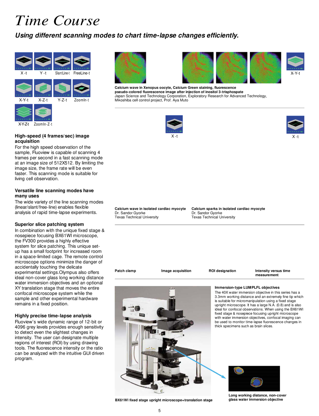
| Calcium wave in Xenopus oocyte, Calcium Green staining, fluorescence | ||
|
|
|
|
|
|
| |||
|
|
|
|
|
|
| |||
|
|
|
|
|
|
| Japan Science and Technology Corporation, Exploratory Research for Advanced Technology, | ||
| |||||||||
| Mikoshiba cell control project, Prof. Aya Muto | ||||||||
|
|
|
|
|
|
|
|
|
|
|
|
|
|
|
|
|
|
|
|
|
|
|
|
|
|
|
|
|
|
High-speed (4 frames/sec) image acquisition
For the high speed observation of the sample, Fluoview is capable of scanning 4 frames per second in a fast scanning mode at an image size of 512X512. By limiting the image size, the frame rate will be even faster. This scanning mode is suitable for living cell observation.
Versatile line scanning modes have many uses
The wide variety of the line scanning modes
X | X |
Calcium wave in isolated cardiac myocyte | Calcium sparks in isolated cardiac myocyte |
Dr. Sandor Gyorke | Dr. Sandor Gyorke |
Texas Technical University | Texas Technical University |
Superior slice patching system
In combination with the unique fixed stage & nosepiece focusing BX61WI microscope, the FV300 provides a highly effective system for slice patching. This unique set- up has a small footprint for increased room in a
Highly precise time-lapse analysis
Fluoview’s wide dynamic range of
Patch clamp | Image acquisition | ROI designation | Intensity versus time |
|
|
| measurement |
|
|
|
|
Immersion-type LUMPLFL objectives
The 40X water immersion objective in this series has a 3.3mm working distance and an extremely fine tip which is suitable for micromanipulation using a fixed stage upright microscope. It has a large N.A. (0.8) and is also ideal for confocal observations. When using the BX61WI fixed stage & nosepiece focusing upright microscope with water immersion objectives, confocal imaging can be used to monitor
| Long working distance, |
BX61WI fixed stage upright microscope+translation stage | glass water immersion objective |
5