Multi-point time lapse system
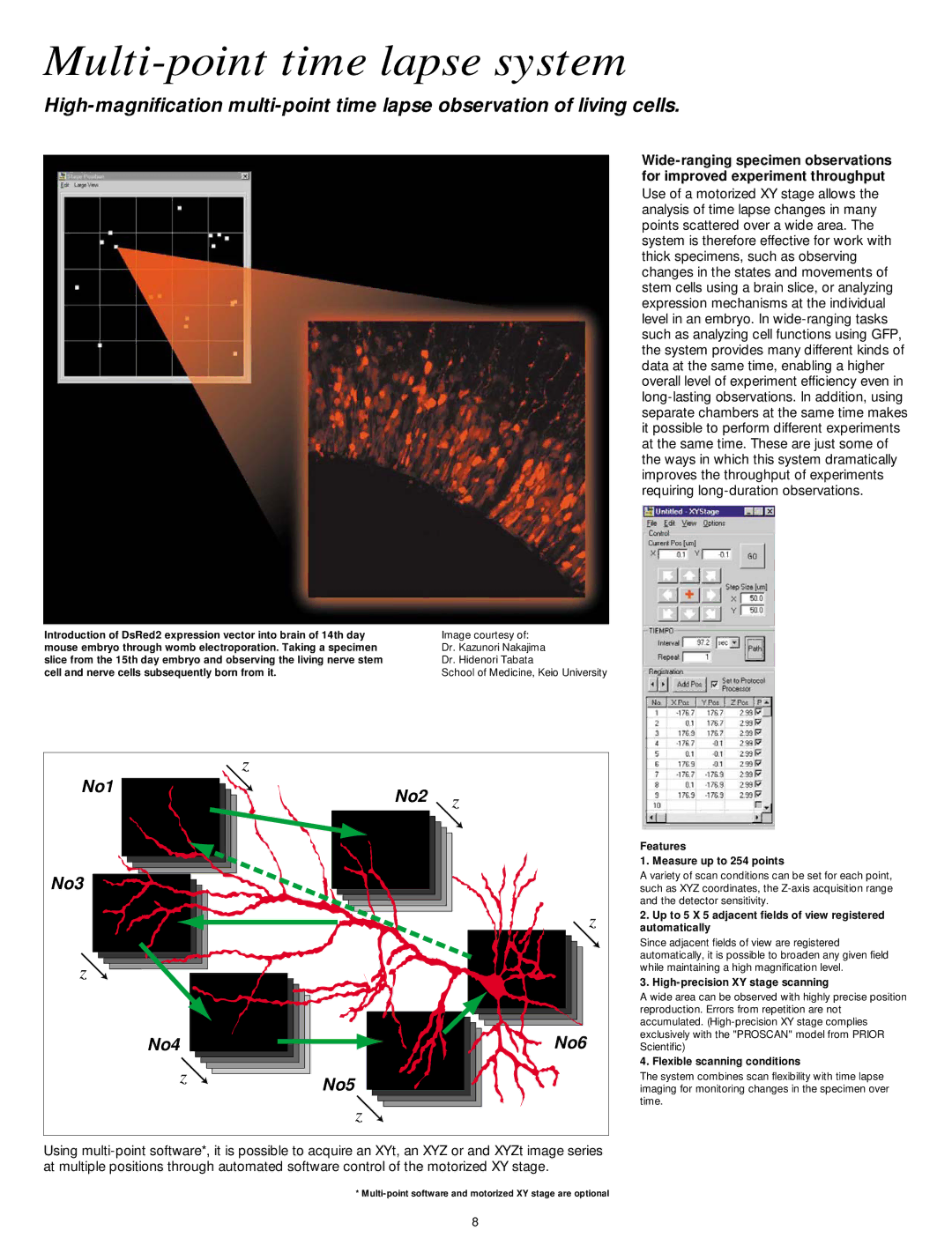
Introduction of DsRed2 expression vector into brain of 14th day mouse embryo through womb electroporation. Taking a specimen slice from the 15th day embryo and observing the living nerve stem cell and nerve cells subsequently born from it.
Use of a motorized XY stage allows the analysis of time lapse changes in many points scattered over a wide area. The system is therefore effective for work with thick specimens, such as observing changes in the states and movements of stem cells using a brain slice, or analyzing expression mechanisms at the individual level in an embryo. In
Image courtesy of:
Dr. Kazunori Nakajima
Dr. Hidenori Tabata
School of Medicine, Keio University
|
| z |
|
No1 |
| No2 | z |
|
| ||
|
|
| |
No3 |
|
|
|
|
|
| z |
z |
|
|
|
No4 |
|
| No6 |
| z | No5 |
|
|
|
| |
|
| z |
|
Using
Features
1. Measure up to 254 points
A variety of scan conditions can be set for each point, such as XYZ coordinates, the
2.Up to 5 X 5 adjacent fields of view registered automatically
Since adjacent fields of view are registered automatically, it is possible to broaden any given field while maintaining a high magnification level.
3.High-precision XY stage scanning
A wide area can be observed with highly precise position reproduction. Errors from repetition are not accumulated.
4. Flexible scanning conditions
The system combines scan flexibility with time lapse imaging for monitoring changes in the specimen over time.
*
8
