OLS3100 specifications
The Olympus OLS3100 is a state-of-the-art laser scanning microscope designed for advanced surface and 3D measurements. It seamlessly combines precision optics with cutting-edge technology, making it suitable for a wide range of applications in materials science, life sciences, and electronics.One of the most notable features of the OLS3100 is its high-resolution imaging capability. Equipped with a robust optical system and advanced laser sources, it enables users to obtain detailed surface and topographical information at nanometer resolution. The microscope employs laser scanning technology that provides rapid data acquisition while minimizing sample damage, which is particularly advantageous when working with delicate specimens.
The OLS3100 also boasts an innovative multi-functional imaging mode. Users can switch between various imaging techniques, such as brightfield, darkfield, and fluorescence, enhancing versatility for different research needs. This multifunctionality allows researchers to study samples from different perspectives, enabling deeper insights into structural and compositional properties.
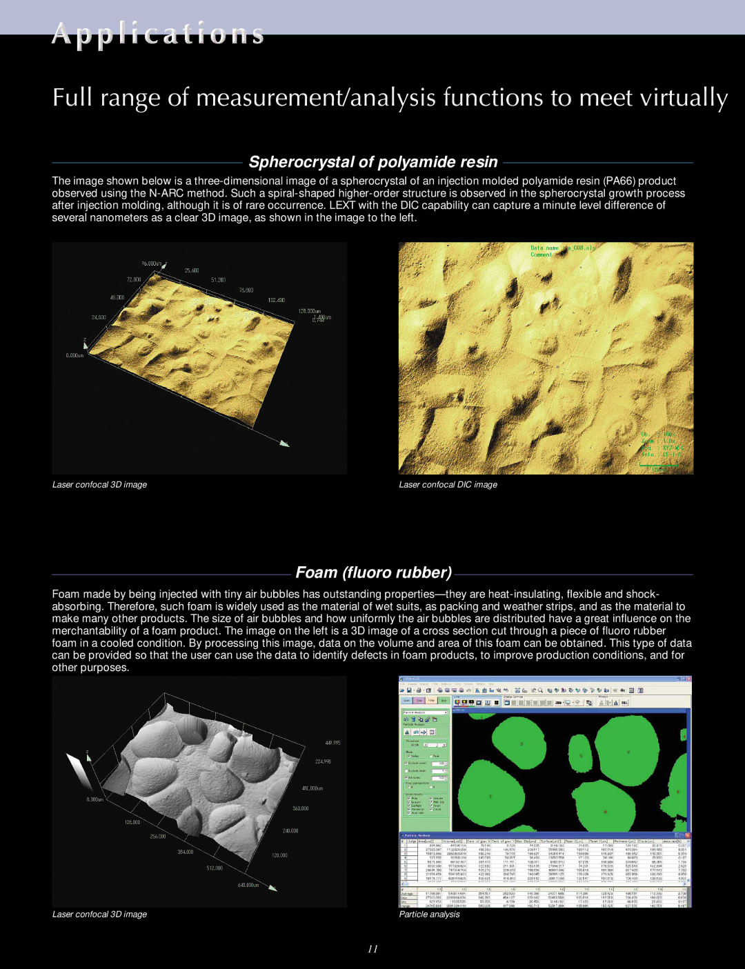
Furthermore, the OLS3100 features advanced software that facilitates easy data analysis and visualization. This software integrates powerful algorithms for surface measurement and metrology, allowing for efficient processing of complex datasets. Users can create 3D reconstructions of the sample surface, conduct roughness analysis, and generate detailed reports effortlessly.
The instrument is designed for user-friendly operation, featuring an intuitive interface and customizable workflows. This enables researchers, regardless of their experience level, to operate the system effectively. The OLS3100 also supports remote access capabilities, making it an ideal choice for collaborative projects where multiple users may need to access and analyze data from different locations.
In terms of durability, the OLS3100 is built with high-quality materials that ensure long-term reliability. The robust construction minimizes vibrations and other environmental interferences, resulting in consistent performance over time.
In conclusion, the Olympus OLS3100 is a versatile and powerful tool that combines high-resolution imaging, multifunctional capabilities, and user-friendly software for comprehensive surface analysis. This advanced laser scanning microscope is an excellent choice for researchers looking to advance their work in various scientific fields while ensuring precision, efficiency, and reliability. Its innovative features make it a valuable asset in any laboratory, pushing the boundaries of what is possible in microscopy today.

