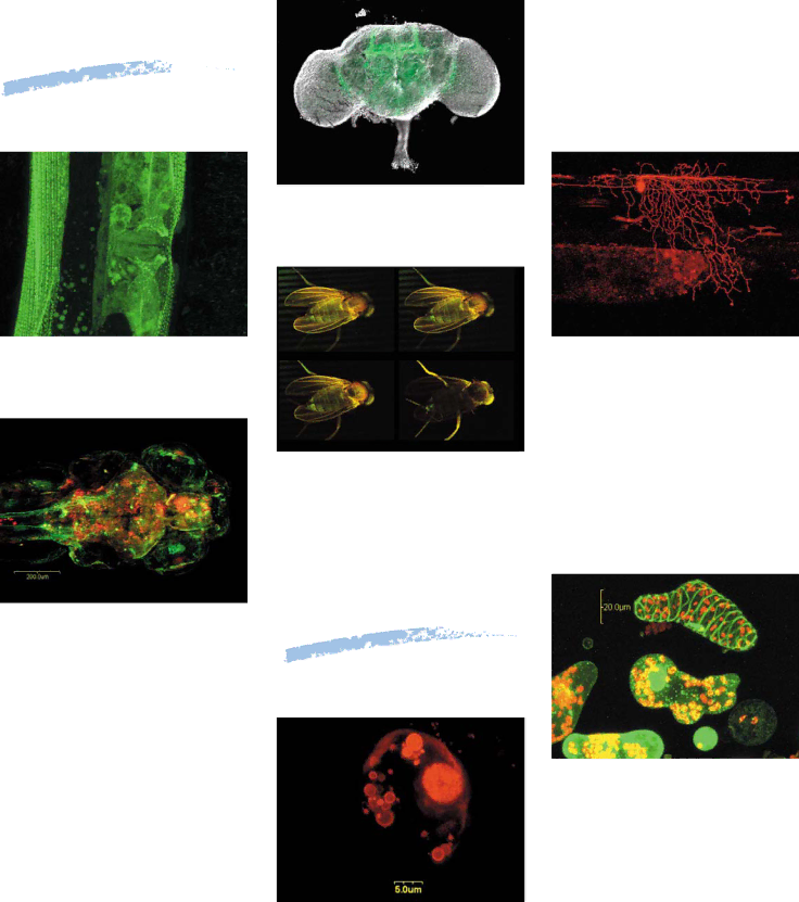
Olympus FV300 manual Fluorescent Proteins

Page 13

Fluorescent

Proteins

GFP-labeling of Drosophila adult brain with staining of mushroom bodies

Assistant Prof. Aigaki

Cytogenetics

Tokyo Metropolitan University, Science Dept.

C elegans expressing beta-integrin fused to GFP

Dr. Xioping Xhu and Dr. John Plenefisch

University of Toledo, Dept. of Biology

GFP-labeling of Drosophila adults

Expression of DsRed in a zebrafish embryo Extended focus image of 5µmx30 slice Pr. Yasuhiro Kamei, Pr. Shunsuke Yuba Institute for Molecular and Cellular Biology Osaka University

Coexpression of EGFP and DsRed in a zebrafish embryo

Extended focus image of 10µmx28 slice Pr. Yasuhiro Kamei, Pr. Shunsuke Yuba Institute for Molecular and Cellular Biology Osaka University

Plant

Isolated Zinnia mesophyll cells

Keisuke Obara

Pr. Hiroo Fukuda

Department of Biological Sciences,

Graduate School of Science,

The University of Tokyo

Apoptosis of Tabacco hybrid plant cells

Dr. Wataru Marubashi

Laboratory of Plant Breeding and Cell Engineering,

School of Agriculture,Ibaraki University

12

Image 13
Contents FV300 FV300-IX71configuration Page Software Graphical User Interface Image tool bar Easy exchange between display methodsThumbnail display Scanning unit set-up monitor displayHigh-speed 4 frames/sec image acquisition Time CourseVersatile line scanning modes have many uses Superior slice patching system440nm diode laser can be added for CFP/YFP Fret imaging Input/output of external trigger signalRatio imaging to analyze 2-wavelength images Papp for Frap Application Multi-point time lapse system Features Measure up to 254 pointsHigh-precision XY stage scanning Flexible scanning conditions3D Imaging Easy Z axis operation and settingRegions/Min-Max Mode Threshold ModeColocalization Link with patch clamping dataStructure of PtK2 cell Applications GalleryFluorescent Proteins Short Pass Band Pass Long Pass 300 400 500 600Fluorochrome Specifications ISO9001