Technology / Hardware
Exceptional Resolution for Imaging of Cytoplasmic Membrane and Areas Deep Within Living Specimen.
TIRFM (Total Internal Reflection Fluorescence Microscope) System
Switchable between Confocal and TIRFM Imaging
Switchable between confocal and TIRFM imaging for localization of proteins on the cytoplasmic membrane surface and acquisition of sectioning images within cells.
Software Control of TIRF Illumination
|
|
|
|
| |||
A line of |
|
|
| ||||
TIRF illumination. |
|
|
|
| TIRFM | LSM | |
|
|
|
|
|
|
| |
|
|
|
|
|
| Courtesy of Dr.J M Dong of | |
NEW | APON60xOTIRF | NEW | UAPON100xOTIRF | NEW | UAPON150xOTIRF | Apo100xOHR | |
| NA : 1.49 (oil immersion) |
| NA : 1.49 (oil immersion) |
| NA : 1.45 (oil immersion) | NA : 1.65 (oil immersion) | |
| WD: 0.1 mm |
| WD: 0.1 mm |
| WD: 0.08 mm | WD: 0.1 mm | |
|
|
|
|
|
|
| (Customized cover glass and |
|
|
|
|
|
|
| immersion oil) |
FV1000MPE Multiphoton Excitation System
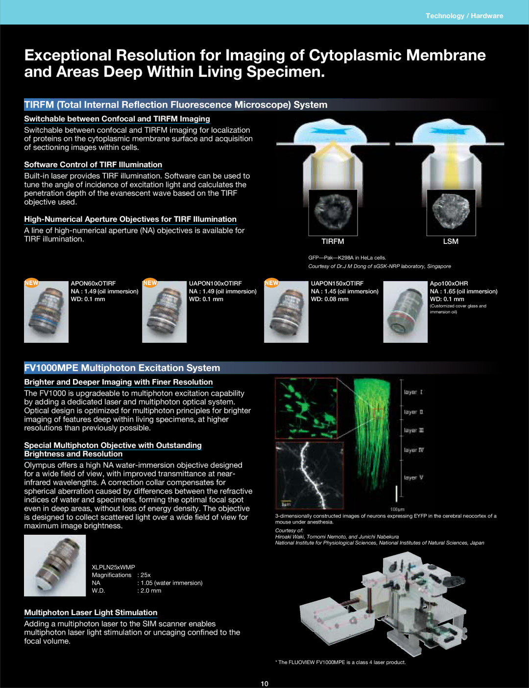
Brighter and Deeper Imaging with Finer Resolution
The FV1000 is upgradeable to multiphoton excitation capability by adding a dedicated laser and multiphoton optical system. Optical design is optimized for multiphoton principles for brighter imaging of features deep within living specimens, at higher resolutions than previously possible.
Special Multiphoton Objective with Outstanding
Brightness and Resolution
Olympus offers a high NA
XLPLN25xWMP |
|
Magnifications | : 25x |
NA | : 1.05 (water immersion) |
W.D. | : 2.0 mm |
Multiphoton Laser Light Stimulation
Adding a multiphoton laser to the SIM scanner enables multiphoton laser light stimulation or uncaging confined to the focal volume.
Courtesy of:
Hiroaki Waki, Tomomi Nemoto, and Junichi Nabekura
National Institute for Physiological Sciences, National Institutes of Natural Sciences, Japan
* The FLUOVIEW FV1000MPE is a class 4 laser product.
10