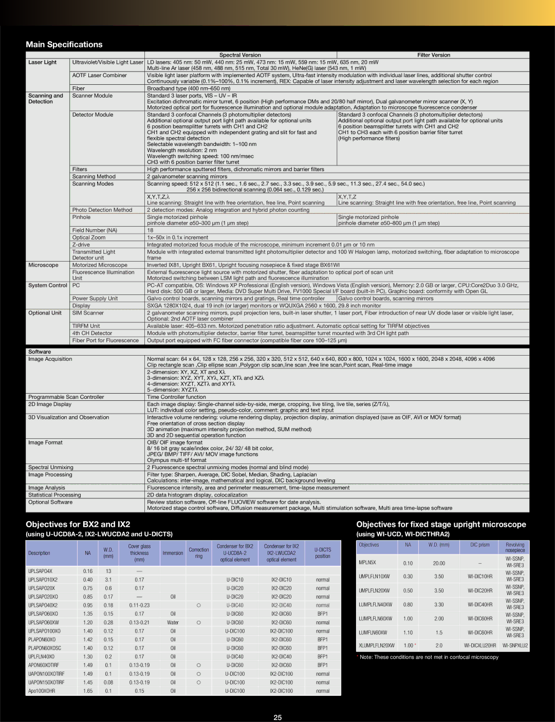
Main Specifications
|
|
| Spectral Version | Filter Version |
Laser Light |
| Ultraviolet/Visible Light Laser | LD lasers: 405 nm: 50 mW, 440 nm: 25 mW, 473 nm: 15 mW, 559 nm: 15 mW, 635 nm, 20 mW | |
|
|
| ||
|
| AOTF Laser Combiner | Visible light laser platform with implemented AOTF system, | |
|
|
| Continuously variable | |
|
| Fiber | Broadband type (400 |
|
Scanning and |
| Scanner Module | Standard 3 laser ports, VIS – UV – IR |
|
Detection |
|
| Excitation dichromatic mirror turret, 6 position (High performance DMs and 20/80 half mirror), Dual galvanometer mirror scanner (X, Y) | |
|
|
| Motorized optical port for fluorescence illumination and optional module adaptation, Adaptation to microscope fluorescence condenser | |
|
| Detector Module | Standard 3 confocal Channels (3 photomultiplier detectors) | Standard 3 confocal Channels (3 photomultiplier detectors) |
|
|
| Additional optional output port light path available for optional units | Additional optional output port light path available for optional units |
|
|
| 6 position beamsplitter turrets with CH1 and CH2 | 6 position beamsplitter turrets with CH1 and CH2 |
|
|
| CH1 and CH2 equipped with independent grating and slit for fast and | CH1 to CH3 each with 6 position barrier filter turret |
|
|
| flexible spectral detection | (High performance filters) |
|
|
| Selectable wavelength bandwidth: |
|
|
|
| Wavelength resolution: 2 nm |
|
|
|
| Wavelength switching speed: 100 nm/msec |
|
|
|
| CH3 with 6 position barrier filter turret |
|
|
| Filters | High performance sputtered filters, dichromatic mirrors and barrier filters |
|
|
| Scanning Method | 2 galvanometer scanning mirrors |
|
|
| Scanning Modes | Scanning speed: 512 x 512 (1.1 sec., 1.6 sec., 2.7 sec., 3.3 sec., 3.9 sec., 5.9 sec., 11.3 sec., 27.4 sec., 54.0 sec.) | |
|
|
| 256 x 256 bidirectional scanning (0.064 sec., 0.129 sec.) |
|
|
|
| X,Y,T,Z,λ | X,Y,T,Z |
|
|
| Line scanning: Straight line with free orientation, free line, Point scanning | Line scanning: Straight line with free orientation, free line, Point scanning |
|
| Photo Detection Method | 2 detection modes: Analog integration and hybrid photon counting |
|
|
| Pinhole | Single motorized pinhole | Single motorized pinhole |
|
|
| pinhole diameter | pinhole diameter |
|
| Field Number (NA) | 18 |
|
|
| Optical Zoom |
| |
|
| Integrated motorized focus module of the microscope, minimum increment 0.01 µm or 10 nm | ||
|
| Transmitted Light | Module with integrated external transmitted light photomultiplier detector and 100 W Halogen lamp, motorized switching, fiber adaptation to microscope | |
|
| Detector unit | frame |
|
Microscope |
| Motorized Microscope | Inverted IX81, Upright BX61, Upright focusing nosepiece & fixed stage BX61WI | |
|
| Fluorescence Illumination | External fluorescence light source with motorized shutter, fiber adaptation to optical port of scan unit | |
|
| Unit | Motorized switching between LSM light path and fluorescence illumination |
|
System Control |
| PC | ||
|
|
| Hard disk: 500 GB or larger, Media: DVD Super Multi Drive, FV1000 Special I/F board | |
|
| Power Supply Unit | Galvo control boards, scanning mirrors and gratings, Real time controller | Galvo control boards, scanning mirrors |
|
| Display | SXGA 1280X1024, dual 19 inch (or larger) monitors or WQUXGA 2560 x 1600, 29.8 inch monitor | |
Optional Unit |
| SIM Scanner | 2 galvanometer scanning mirrors, pupil projection lens, | |
|
|
| Optional: 2nd AOTF laser combiner |
|
|
| TIRFM Unit | Available laser: | |
|
| 4th CH Detector | Module with photomultiplier detector, barrier filter turret, beamsplitter turret mounted with 3rd CH light path | |
|
| Fiber Port for Fluorescence | Output port equipped with FC fiber connector (compatible fiber core | |
|
|
|
|
|
Software |
|
|
| |
Image Acquisition |
| Normal scan: 64 x 64, 128 x 128, 256 x 256, 320 x 320, 512 x 512, 640 x 640, 800 x 800, 1024 x 1024, 1600 x 1600, 2048 x 2048, 4096 x 4096 | ||
|
|
| Clip rectangle scan ,Clip ellipse scan ,Polygon clip scan,line scan ,free line scan,Point scan, | |
|
|
|
| |
|
|
|
| |
|
|
|
| |
|
|
| 5−dimension: XYZTλ |
|
Programmable Scan Controller | Time Controller function |
| ||
2D Image Display |
| Each image display: | ||
|
|
| LUT: individual color setting, |
|
3D Visualization and Observation | Interactive volume rendering: volume rendering display, projection display, animation displayed (save as OIF, AVI or MOV format) | |||
|
|
| Free orientation of cross section display |
|
|
|
| 3D animation (maximum intensity projection method, SUM method) |
|
|
|
| 3D and 2D sequential operation function |
|
Image Format |
| OIB/ OIF image format |
| |
|
|
| 8/ 16 bit gray scale/index color, 24/ 32/ 48 bit color, |
|
|
|
| JPEG/ BMP/ TIFF/ AVI/ MOV image functions |
|
|
|
| Olympus |
|
Spectral Unmixing | 2 Fluorescence spectral unmixing modes (normal and blind mode) |
| ||
Image Processing |
| Filter type: Sharpen, Average, DIC Sobel, Median, Shading, Laplacian |
| |
|
|
| Calculations: |
|
Image Analysis |
| Fluorescence intensity, area and perimeter measurement, | ||
Statistical Processing | 2D data histogram display, colocalization |
| ||
Optional Software | Review station software, |
| ||
|
|
| Motorized stage control software, Diffusion measurement package, Multi stimulation software, Multi area | |
|
|
|
|
|
Objectives for BX2 and IX2
(using U-UCD8A-2, IX2-LWUCDA2 and U-DICTS)
|
| W.D. | Cover glass |
| Correction | Condenser for BX2 | Condenser for IX2 | ||
Description | NA | thickness | Immersion | ||||||
(mm) | ring | position | |||||||
|
| (mm) |
| optical element | optical element | ||||
|
|
|
|
|
| ||||
UPLSAPO4X | 0.16 | 13 | — |
|
|
|
|
| |
UPLSAPO10X2 | 0.40 | 3.1 | 0.17 |
|
| normal | |||
UPLSAPO20X | 0.75 | 0.6 | 0.17 |
|
| normal | |||
UPLSAPO20XO | 0.85 | 0.17 | — | Oil |
| normal | |||
UPLSAPO40X2 | 0.95 | 0.18 |
| _ | normal | ||||
UPLSAPO60XO | 1.35 | 0.15 | 0.17 | Oil |
| BFP1 | |||
UPLSAPO60XW | 1.20 | 0.28 | Water | _ | normal | ||||
UPLSAPO100XO | 1.40 | 0.12 | 0.17 | Oil |
| normal | |||
PLAPON60XO | 1.42 | 0.15 | 0.17 | Oil |
| BFP1 | |||
PLAPON60XOSC | 1.40 | 0.12 | 0.17 | Oil |
| BFP1 | |||
UPLFLN40XO | 1.30 | 0.2 | 0.17 | Oil |
| BFP1 | |||
APON60XOTIRF | 1.49 | 0.1 | Oil | _ | BFP1 | ||||
UAPON100XOTIRF | 1.49 | 0.1 | Oil | _ | normal | ||||
UAPON150XOTIRF | 1.45 | 0.08 | Oil | _ | normal | ||||
Apo100XOHR | 1.65 | 0.1 | 0.15 | Oil |
| normal |
Objectives for fixed stage upright microscope
(using WI-UCD, WI-DICTHRA2)
Objectives | NA | W.D. (mm) | DIC prism | Revolving | |
|
|
|
| nosepiece | |
MPLN5X | 0.10 | 20.00 | – | ||
|
|
|
| ||
UMPLFLN10XW | 0.30 | 3.50 | |||
|
|
|
| ||
UMPLFLN20XW | 0.50 | 3.50 | |||
|
|
|
| ||
LUMPLFLN40XW | 0.80 | 3.30 | |||
|
|
|
| ||
LUMPLFLN60XW | 1.00 | 2.00 | |||
|
|
|
| ||
LUMFLN60XW | 1.10 | 1.5 | |||
|
|
|
| ||
XLUMPLFLN20XW | 1.00 * | 2.0 | |||
|
|
|
|
|
*Note: These conditions are not met in confocal microscopy
25
