Application/ Molecular Interaction Analysis
Photoconversion
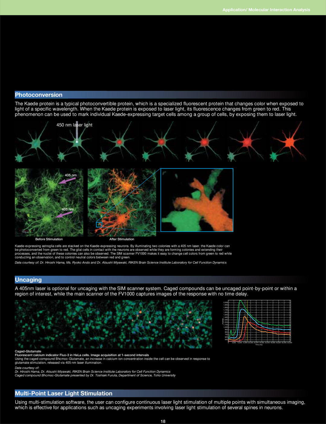
The Kaede protein is a typical photoconvertible protein, which is a specialized fluorescent protein that changes color when exposed to light of a specific wavelength. When the Kaede protein is exposed to laser light, its fluorescence changes from green to red. This phenomenon can be used to mark individual Kaede-expressing target cells among a group of cells, by exposing them to laser light.
450 nm laser light
405 nm
405 nm
Before Stimulation | After Stimulation |
Kaede-expressing astroglia cells are stacked on the Kaede-expressing neurons. By illuminating two colonies with a 405 nm laser, the Kaede color can be photoconverted from green to red. The glial cells in contact with the neurons are observed while they are forming colonies and extending their processes, and the nuclei of these colonies can also be observed. The SIM scanner FV1000 makes it easy to change cell colors from green to red while conducting an observation, and to control neutral colors between red and green.
Data courtesy of: Dr. Hiroshi Hama, Ms. Ryoko Ando and Dr. Atsushi Miyawaki, RIKEN Brain Science Institute Laboratory for Cell Function Dynamics
Uncaging
A 405nm laser is optional for uncaging with the SIM scanner system. Caged compounds can be uncaged point-by-point or within a region of interest, while the main scanner of the FV1000 captures images of the response with no time delay.
2,000
1,900
1,800
1,700
1,600
1,500
1,400
1,300
1,200
1,100
1,000
900
800
700
600
500
0 5,000 10,000 15,000 20,000 25,000 30,000 35,000 40,000 45,000 50,000 55,000 Time (ms)
Caged-Glutamate
Fluorescent calcium indicator Fluo-3 in HeLa cells. Image acquisition at 1-second intervals
Using the caged compound Bhcmoc-Glutamate, an increase in calcium ion concentration inside the cell can be observed in response to glutamate stimulation, released via 405 nm laser illumination.
Data courtesy of:
Dr. Hiroshi Hama, Dr. Atsushi Miyawaki, RIKEN Brain Science Institute Laboratory for Cell Function Dynamics
Caged compound Bhcmoc-Glutamate presented by Dr. Toshiaki Furuta, Department of Science, Toho University
Multi-Point Laser Light Stimulation
Using multi-stimulation software, the user can configure continuous laser light stimulation of multiple points with simultaneous imaging, which is effective for applications such as uncaging experiments involving laser light stimulation of several spines in neurons.

