9. Kontrastverfahren für Leica DM4000 B/DM4500 P/DM5000 B

Einfach-Übersichtsbeobachtung

Legen Sie ein Durchlichtpräparat auf den Polarisator auf.

Schwenken Sie den Kondensorkopf ein.

Fokussieren Sie mit einem schwach ver- größernden Objektiv, z. B. 5x, durch den Kon- densor.

Wenngleich diese Methode keinen Anspruch auf gute Abbildungsleistung hat, so ermöglicht sie z. B. sehr schnelles Sichten von Präparat- reihen.

λ/4- undλ-Platte, Quarzkeil

λ/4- und λ-Platte werden je nach Ausführung an der Kondensorunterseite oder bei Polarisations- mikroskopen in die Kondensorscheibe 8fach eingebaut (die Schwingungsrichtung γ ver-

läuft: ) oder in den Tubusschlitz eingesteckt. Der Tubusschlitz ist durch eine selbsttätig fe- dernde Staubschutzklappe verschlossen.

Beim Analysator IC/P kann durch Wenden, so dass die Gravur l nach oben zeigt, die λ-Platte aktiviert werden.

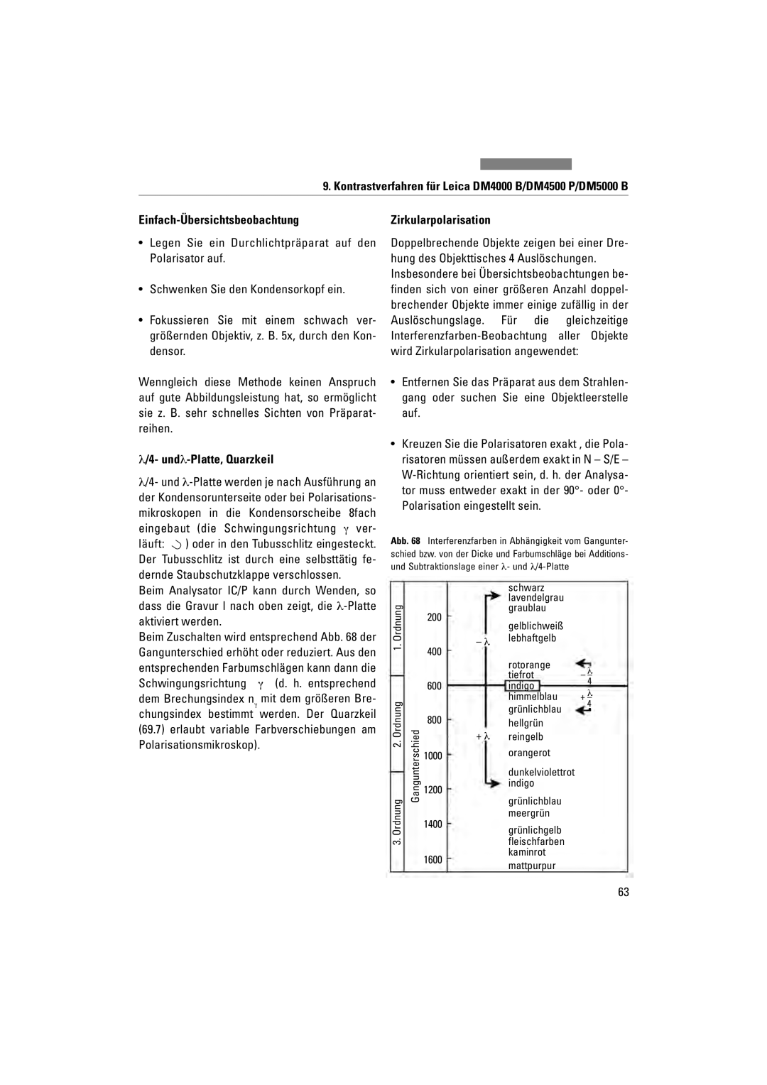
Beim Zuschalten wird entsprechend Abb. 68 der Gangunterschied erhöht oder reduziert. Aus den entsprechenden Farbumschlägen kann dann die Schwingungsrichtung γ (d. h. entsprechend dem Brechungsindex nγ mit dem größeren Bre- chungsindex bestimmt werden. Der Quarzkeil (69.7) erlaubt variable Farbverschiebungen am Polarisationsmikroskop).

Zirkularpolarisation

Doppelbrechende Objekte zeigen bei einer Dre- hung des Objekttisches 4 Auslöschungen. Insbesondere bei Übersichtsbeobachtungen be- finden sich von einer größeren Anzahl doppel- brechender Objekte immer einige zufällig in der Auslöschungslage. Für die gleichzeitige Interferenzfarben-Beobachtung aller Objekte wird Zirkularpolarisation angewendet:

Entfernen Sie das Präparat aus dem Strahlen- gang oder suchen Sie eine Objektleerstelle auf.

Kreuzen Sie die Polarisatoren exakt , die Pola- risatoren müssen außerdem exakt in N – S/E – W-Richtung orientiert sein, d. h. der Analysa- tor muss entweder exakt in der 90°- oder 0°- Polarisation eingestellt sein.

Abb. 68 Interferenzfarben in Abhängigkeit vom Gangunter- schied bzw. von der Dicke und Farbumschläge bei Additions- und Subtraktionslage einer λ- und λ/4-Platte


schwarz


lavendelgrau


Ordnung


200

graublau


lebhaftgelb


gelblichweiß


1.


λ


400


rotorange

λ


tiefrot

– –


600

indigo

4


himmelblau

λ


+ –

Ordnung3. 2. Ordnung


grünlichblau

4


800


Gangunterschied

hellgrün


+ λ

fleischfarben


reingelb


1000

orangerot


dunkelviolettrot


1200

indigo

grünlichblau


1400

meergrün

grünlichgelb


1600

kaminrot

mattpurpur


63

Page 143
Image 143
Leica DM4500P, DM5000B, DM4000M manual Einfach-Übersichtsbeobachtung, Undλ-Platte, Quarzkeil, Zirkularpolarisation

DM4500P, DM5000B, DM4000M specifications

The Leica DM4000M and DM4000B are state-of-the-art microscopes designed for professional use in the fields of biology, materials science, and clinical applications. Renowned for their precision and innovative features, these instruments are perfect for researchers and clinicians needing high-resolution imaging capabilities.

One of the standout features of both models is the advanced motorized focusing system, which allows for swift adjustments and precise control. This feature is particularly useful in time-sensitive research environments, where accuracy and speed are paramount. The ergonomically designed focus mechanism promotes user comfort during prolonged observation sessions.

Both the DM4000M and DM4000B incorporate the revolutionary Leica Application Suite (LAS) software. This intuitive platform is designed to maximize the functionality of the microscope, enabling users to capture, analyze, and share images seamlessly. The software’s integrated tools are perfect for documenting findings and enhancing research productivity.

Another notable characteristic of the DM4000 series is the modular design, which allows for easy customization and upgrading. This aspect ensures that users can tailor their microscopes to meet specific research needs, whether it be for fluorescence microscopy, phase contrast, or even special imaging techniques like HSR or IR.

The high-performance optics provide exceptional image contrast and clarity, allowing users to observe minute details in samples. The combination of high numerical aperture objectives and advanced optical coatings enhances the resolution, making the DM4000 series ideal for examining intricate biological specimens as well as materials with complex textures.

The DM4000B model is particularly suited for routine clinical applications, featuring specific tools designed for rapid diagnosis and efficient workflows. Its user-friendly interface and dedicated clinical applications streamline laboratory processes, making it an essential device in any clinical setting.

Additionally, both models are equipped with LED illumination, which offers consistent light intensity and color temperature. This feature improves sample clarity while reducing heat generation, thereby protecting sensitive specimens during prolonged observation periods.

In conclusion, the Leica DM4000M and DM4000B microscopes represent a blend of advanced technology, intuitive design, and high adaptability. Their user-focused features and exceptional optical performance make them indispensable tools for researchers and clinical professionals aiming for excellence in microscopy.