syngo Osteo CT
•Position the cut line of scanning through the middle of the vertebra, i.e.
•The phantom must be included in the FoV of the images for evaluation.
•It is recommended to end the exam first, and then start the syngo Osteo CT evaluation.
•Do not use the calibration phantom from other CT scanners, as your system is calibrated to a particular phantom.
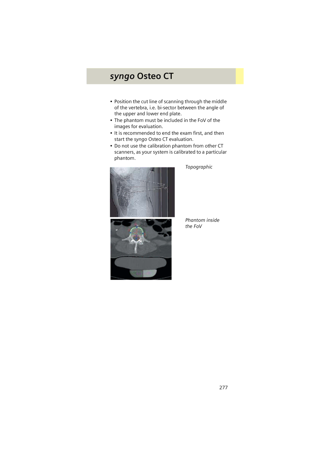
Topographic
Phantom inside the FoV
277