syngo Volume Evaluation
Workflow
1. Loading the Images
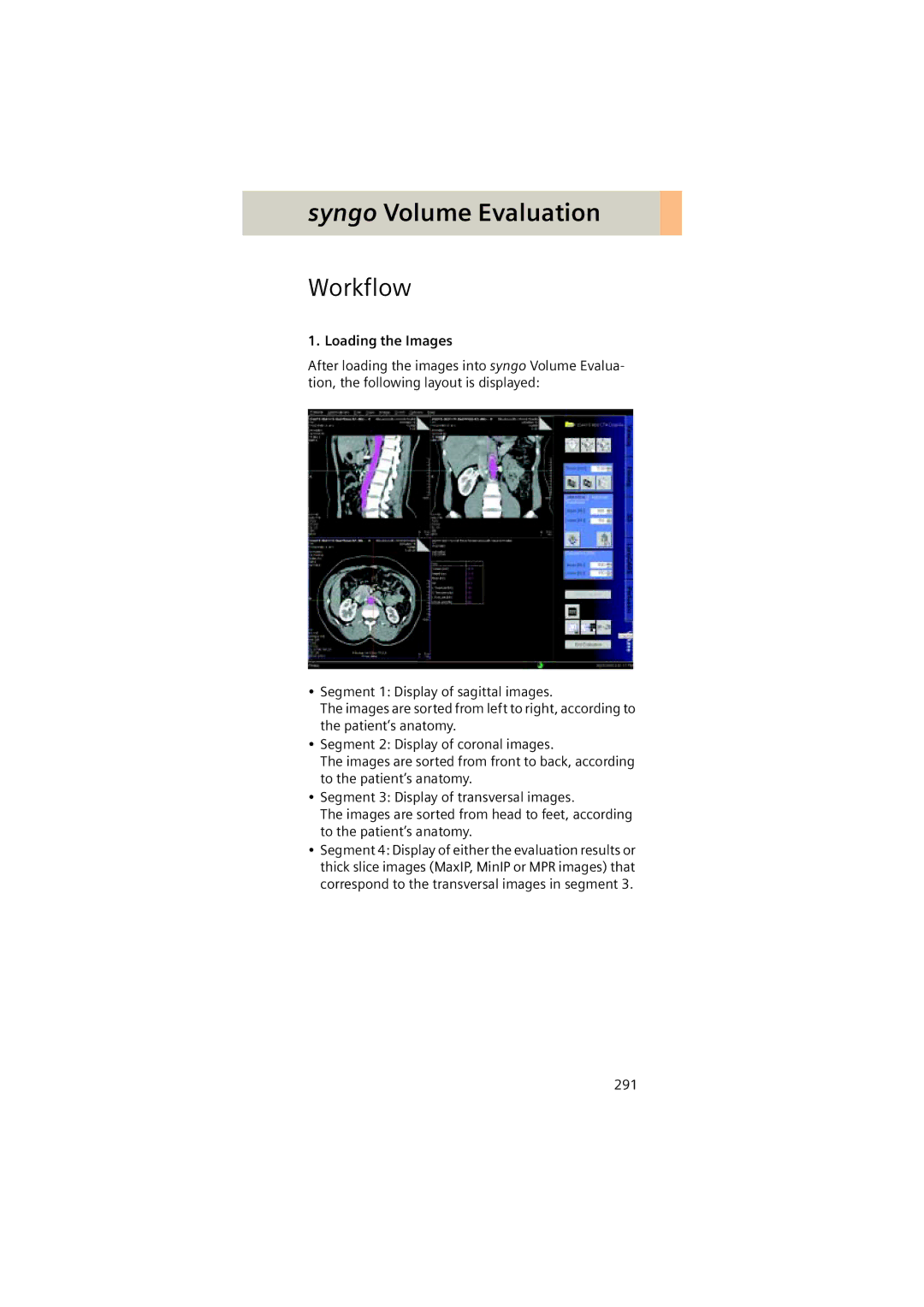
After loading the images into syngo Volume Evalua- tion, the following layout is displayed:
•Segment 1: Display of sagittal images.
The images are sorted from left to right, according to the patient’s anatomy.
•Segment 2: Display of coronal images.
The images are sorted from front to back, according to the patient’s anatomy.
•Segment 3: Display of transversal images.
The images are sorted from head to feet, according to the patient’s anatomy.
•Segment 4: Display of either the evaluation results or thick slice images (MaxIP, MinIP or MPR images) that correspond to the transversal images in segment 3.
291