3.5Operation for Image Observation
Follow the operation below for observation of a scanning image.
• | Selecting Magnification | (3.1.1) |
• | Selecting Scanning Speed | (3.1.2) |
• | Image Brightness and Contrast Adjustment | (3.1.3) |
• | Focus Adjustment and Astigmatism Correction..(3.1.3) | |
• | Operation of Specimen Stage Type I |
|
| (Manual Stage) | (3.1.3) |
• | Operation of Specimen Stage Type II |
|
| (3.1.3) | |
|
|
|
3.5.1SE detector
The upper secondary electron detector is placed above the objective lens and secondary electrons are detected through the magnetic field of the lens. The lower secondary electron detector is placed in the specimen chamber, where a large amount of the signal is due to backscattered electrons. Signals from these two detectors can be selected individually, or mixed together. The image contrast of these detector signals is unique in that each has its own special characteristics.
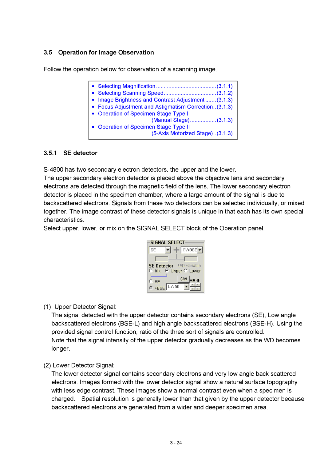
Select upper, lower, or mix on the SIGNAL SELECT block of the Operation panel.
(1) Upper Detector Signal:
The signal detected with the upper detector contains secondary electrons (SE), Low angle backscattered electrons
Note that the signal intensity of the upper detector gradually decreases as the WD becomes longer.
(2) Lower Detector Signal:
The lower detector signal contains secondary electrons and very low angle back scattered electrons. Images formed with the lower detector signal show a natural surface topography with less edge contrast. These images show a normal contrast even when a specimen is charged. Spatial resolution is generally lower than that given by the upper detector because backscattered electrons are generated from a wider and deeper specimen area.
3 - 24
